What is the placenta?
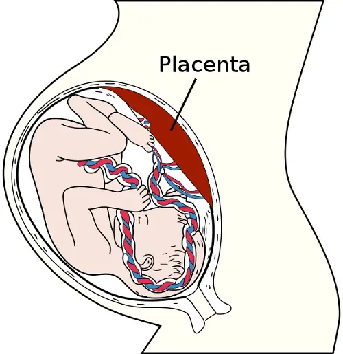
The placenta is a disc shaped organ that has a weight of about 500 g and diameter of 185 mm. It is attached to the fetus via the umbilical cord and to the endometrium of the uterus.
The connection to the fetus is via the umbilical cord which connects to the placenta. At implantation, the placenta begins to develop from the blastocyst of the fetus and deciduo basalis of the endometrium.
The placenta is partly formed from embryonic tissues of the syncytiotrophoblast, cytotrophoblast and mesoderm. These form the chorionic membrane, which further proliferates into the chorionic villi, which form lobules on the maternal side. The villi are folded structures which greatly increase the surface area for absorption of substances, and are surrounded by intervillous spaces.
The cytotrophoblast cells of the trophoblast modify the mother’s blood vessels to supplement blood flow entering the placenta while, the fetal mesenchyme cells form the blood vessels in the chorionic villi.
The placenta takes over the function of feeding the embryo at weeks 4 to 12 of development. The chorionic villi develops simultaneously with the fetus as it grows, becoming more branched and thin-walled, increasing the rate of gas exchange.
Blood from the mother flows into the intervillous spaces while blood from the fetus flows through capillaries that enter the villi. Nutrients and gases are then exchanged between mother and fetus by moving through the membranous walls of the villi.
- Carbon dioxide, oxygen and lipid-soluble substances move across the membrane of the villi by simple diffusion.
- Glucose moves across to the fetus by facilitated diffusion while,
- iron and amino acids move across by active transport.
Blood from mother and fetus don’t mix to prevent the maternal cytotoxic T cells from attacking the fetus. This is because the mother’s antigens would be recognized as non-self.
Nutrients and oxygen from the mother’s blood move into the fetal circulation while the waste products of urea, creatinine and carbon dioxide pass from the fetus into the mother’s blood.
The IgG antibodies from the mother pass to the fetus and confer passive immunity on it. The placenta does act as a barrier to prevent some microbes from passing from mother to fetus, but some harmful substances like alcohol can pass through.
The placenta produces the hormones progesterone and estrogen that control pregnancy, along with several other hormones such as human chorionic gonadotropin, ACTH, human placental lactogen, and parathyroid hormone.
What is the umbilical cord?
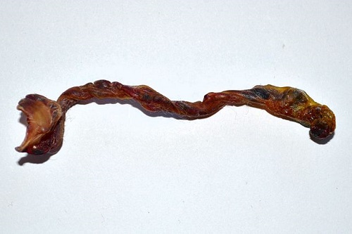
The umbilical cord is elongated and varies in length from 30 to 80 cm, with varying thickness along its length. The cord attaches to the fetus at the naval and attaches near the center of the chorionic plate of the placenta.
It forms from the connecting stalk that was derived from the vitelline duct. The outermost layer of the umbilical cord is produced from the allantois and yolk sac. This layer of the umbilical cord consists of chorionic epithelium, while the inside of the cord contains two arteries and one vein.
The cord is completely formed by the fifth week of development and functions in housing and protecting the blood vessels. These blood vessels are surrounded by Wharton’s jelly, a mucous connective tissue which forms from the mesenchyme cells during development and resists twisting and compression, hence acting as a cushion to protect the blood vessels.
Where the umbilical cord attaches to the mother’s placenta the arteries and vein further divide to become capillaries that enter the chorionic villi. The umbilical arteries carry deoxygenated fetal blood and wastes back to the mother, while the one umbilical vein carries oxygenated blood and nutrients from the mother to the fetus.
Oxygenated blood passes from the umbilical vein into the inferior vena cava of the fetus. From there it passes to the ductus venosus of the heart. The arteries carry the waste products such as urea, in addition to carbon dioxide, while the vein carries oxygen, hormones, and nutrients to the fetus.
These umbilical blood vessels are formed during development from extra embryonic mesodermal tissue. The umbilical arteries are formed from the iliac arteries of the fetus. Initially there are two veins, but the right umbilical vein disappears by the fourth month of development.
What is the difference between the placenta and umbilical cord?
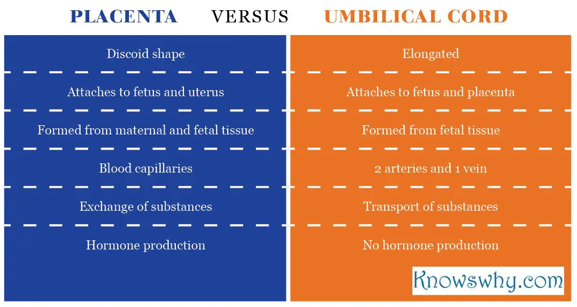
- The placenta is discoid in shape, and attaches to the wall of the uterus whereas, the umbilical cord is elongated and attaches to the placenta and to the fetus.
- Only the umbilical cord is directly attached to the fetus at the umbilical region (naval), the placenta is indirectly attached via the umbilical cord.
- While the placenta is formed from both maternal endometrial tissue and fetal embryonic tissues, the umbilical cord is formed from fetal tissues alone.
- The placenta contains numerous blood capillaries, while the umbilical cord only contains two arteries and one vein.
- The placenta is the site at which exchange of substances occur between the blood of mother and fetus, while the cord simply transports blood to and from the fetus.
- The umbilical cord does not play a role in hormone production, whilst the placenta does.
Table comparing the placenta and umbilical cord
Summary:
- The placenta and umbilical cord both form during pregnancy at different times.
- The placenta is discoid shaped while umbilical cord is elongated.
- The placenta develops from the embryo and uterus. The cord develops from fetal tissue.
- Blood capillaries of the fetus and blood vessels from the mother occur in the placenta, but the blood stays separated.
- The umbilical cord contains two arteries and one vein.
- The placenta and umbilical cord both ensure that blood containing gases, nutrients and wastes is circulated between fetus and mother.
- Hormones are produced from the placenta but not the umbilical cord.
Author: Dr. Rae Osborn
Dr. Rae Osborn holds Honours Bachelor of Science degrees in Zoology and Entomology, and Masters of Science in Entomology from the University of Natal in South Africa. She has received a PhD in Quantitative Biology from the University of Texas at Arlington. She was a tenured Associate Professor of Biology at Northwestern State University in Louisiana for 10 years. She also completed an AAS Degree in Information Network Specialist and an AAS in Computer Information Systems, at Bossier Parish Community College in Louisiana.


Leave a Reply