Introduction
Inhalation and exhalation are automatic functions controlled by the autonomic nervous system [1]. The act of breathing oxygenates the blood and removes carbon dioxide, a metabolic waste product.
Lungs
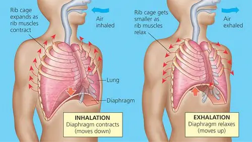
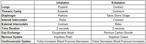
The lungs are made of elastic tissue that expands and contracts during breathing. The lungs do not contain muscle [1]. During inhalation the lungs expand to accommodate air intake. During exhalation the lungs contract as air is expelled.
Thoracic Cavity
The thoracic cavity is the chamber that houses and protects the lungs in addition to other organs, tissues, nerves and cardiovascular vessels. During inhalation the thoracic cavity expands. During exhalation the thoracic cavity contracts.
Diaphragm
The diaphragm is a C-shaped layer of muscle and tissue separating the lungs from the abdomen [1]. The diaphragm is also shaped like a dome. During inhalation, the diaphragm contracts and moves downward, temporarily abandoning the dome-like resting shape. This flattening of the diaphragm enables expansion of the lungs [1]. During exhalation the diaphragm relaxes and moves upward, increasing its dome-like shape to assist with the expulsion of air from the lungs. Relaxation of the diaphragm during exhalation is a passive process that requires no effort from the body [1]. Relaxation of the diaphragm is controlled by nerve signals sent from the vagus and phrenic nerves [1].
i) Inhalation
During inhalation the diaphragm muscles contract and the diaphragm flattens.
ii) Exhalation
During exhalation the diaphragm muscles relax and the diaphragm takes on a dome shape.
Intercostal Muscles
The regions between rib cage bones are filled with intercostal muscles [1] These muscles aid in the inhalation and expulsion of air during respiration. The intercostal muscles originate on ribs 1-11 and are inserted on ribs 2-12 [1]. The intercostal muscles are further divided into external intercostals, internal intercostals and innermost intercostal muscles [1].
The external intercostal muscles enable the elevation of the rib cage and expand the thoracic cavity during respiration by bending the ribs as the muscles contract [1]. The internal intercostal muscles assist in the depression of the rib cage during respiration [1]. Contraction of the internal intercostal muscles helps bend the ribs inward which decreases the volume of the thoracic cavity [1]. The innermost intercostal muscles are a deep layer of muscle separated from the internal intercostal muscles by a neurovascular bundle of nerves and blood vessels [1]. The innermost intercostal muscles assist in exhalation by reducing the volume of the thoracic cavity with their contractions [1].
i) Inhalation
During inhalation the external intercostal muscles contract and the internal intercostal muscles relax.
ii) Exhalation
During exhalation the external intercostal muscles relax and the external intercostal muscles contract
Time Duration of Breathing
Inhalation and exhalation occur for different time periods. The average inhalation takes two seconds [1]. The average exhalation takes three seconds [1].
Respiratory Gas Exchange
Gas exchange during respiration is a passive process by which oxygen and carbon dioxide cross the pulmonary capillaries in the alveolar lung tissue [1]. In gas exchange, oxygen and carbon dioxide diffuse across alveolar surface membranes. During inhalation, oxygen enters the bloodstream. During exhalation carbon dioxide exits the bloodstream.
The thinness of the alveolar membrane enables passive diffusion of oxygen and carbon dioxide. Passive diffusion of the gases is achieved by the pressure differentials between pulmonary capillaries and the alveolar sacs [1]. The pressure of gases in the blood will equilibrate with the alveoli so that oxygen tension maintains a pressure of 100 mmHg and carbon dioxide tension maintains a pressure of 40 mmHg in both arteries and pulmonary capillaries [1]. Inhalation and exhalation rates are adjusted by the body to maintain this pressure throughout the entire circulatory system [1]. This continuous adjustment is an automatic process controlled by the the autonomic nervous system.
i) Inhalation
During inhalation Oxygen crosses the alveolar membrane into the bloodstream during.
ii) Exhalation
During exhalation carbon dioxide is expelled out of the blood stream, across the alveolar membrane into the lungs where it is then pushed out of the body.
Nervous System
Respiratory Centers Monitor Blood pH
Breathing is largely an involuntary action controlled by the upper brainstem where systems called respiratory centers are found [1]. These respiratory regions utilize receptors that continuously monitor blood pH levels [1]. As the blood becomes too acidic, signals are sent throughout the respiratory center that initiate an increase in respiration [1]. This occurs because exhalation essentially removes carbon dioxide from the blood, which raises the pH of blood. Raising bloodstream pH avoids acidosis of the blood.
The medulla oblongata of the upper brainstem is home to the primary respiratory center while aortic bodies located in near the heart and carotid bodies located in the throat serve as peripheral sensors outside of the brain to assist in this continuous monitoring [1]. The respiratory control center sends signals through the nervous system that initiate the contractions and relaxation of intercostal muscles resulting in both inhalations and exhalation [1].
The respiratory center is subdivided into a ventral respiratory control group and a dorsal respiratory control group [1]. Each center performs related but discrete functions that contribute to inhalation and exhalation. The ventral respiratory control group sends nerve impulses through the phrenic nerve to the diaphragm during inhalation [1]. These impulses cause diaphragm contraction and flattening. The dorsal respiratory control group attenuates the rate of breathing and maintains 12 to 16 breaths per minute [1].
i) Inhalation
Initiated by nerve signals sent from the respiratory center.
ii) Exhalation
Initiated by termination of nerve impulses sent from the respiratory center.
Apneustic & Pneumotaxic Centers
Underneath the medulla oblongata is the pons, an additional respiratory center contributing impulses that modify and control the rate of breathing [1]. This respiratory center is further subdivided into two systems, the apneustic center and the pneumotaxic center [1]. The apneustic center sends signals to stimulate long deep inhalations [1]. The pneumotaxic center sends signals that are antagonistic to those sent by the apneustic center and, in doing so, attenuates the rate of breathing [1].
i) Inhalation
The apneustic center primarily stimulates inhalation.
ii) Exhalation
The pneumotaxic center limits inhalation.
Cardiovascular System
The heart exhibits a naturally occurring variation in heart beats that correlates to the acts of inhalation and exhalation [1]. Heart rate increases during inhalation and decreases during exhalation [1]. This phenomenon is called respiratory sinus arrhythmia and is controlled by the vagus nerve [1]. Blood pressure slightly declines during inhalation and increases during exhalation [1]. The normal change in blood pressure between inhalation and exhalation is 10 mmHg or less [1]. As the thoracic cavity expands during inhalation the pressure within it becomes more negative relative to atmospheric pressure [1]. This shift in pressure allows blood vessel expansion and blood rushes into the thoracic vasculature where it becomes oxygenated [1].
Summary


Leave a Reply