The right diagnosis, a crucial component of efficient treatment, can be reached with the help of the appropriate imaging. Therefore, it’s critical to select an imaging facility with a wide range of technology, as well as radiologists and technologists with experience and training in particular body parts, diseases, and imaging techniques.
Doctors frequently use diagnostic imaging procedures to narrow down potential causes of pain or sickness for a more precise diagnosis. The kind of imaging used depends on the region of the body the doctor wants to see on an image and the type of imaging readily available to the patient.
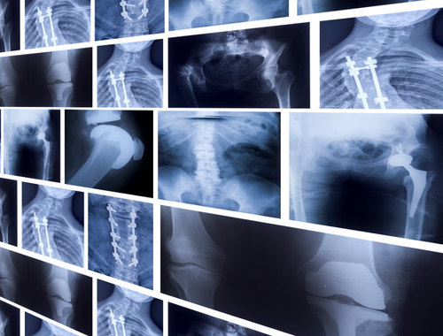
What are X-rays?
A radiograph, often known as an X-ray, emits radiation that travels through the body. Bones and teeth are tissues with significant calcium content that block radiation and look white in images. Additionally, tissues allow radiation to flow through, resulting in a gray or black appearance on the image.
X-ray is the quickest and easiest imaging method and only takes minutes to complete. However, an X-ray cannot show a small bone fracture, soft tissue damage, or inflammation. Therefore, even if your doctor may be concerned about a soft tissue injury, such as a tendon tear, they frequently have an X-ray test to rule out a fracture.
X-rays are frequently used to examine and diagnose cancers, infections, degenerative bone disease, fractures, and dislocations. In addition, an X-ray can be used to check inside structures such as organs in addition to the skeletal features that are frequently examined.
The doctor may give the patient barium sulfate or a dye to make the organs more visible in the X-ray image. The body portion being examined is positioned between an X-ray machine and photographic film in an X-ray.
The patient’s internal structures are then reflected on the exposed film when electromagnetic waves (radiation) travel through the patient’s body. Even though the radiation used for an X-ray is not regarded as harmful, if the patient is pregnant, doctors will take further care.
What are MRIs?
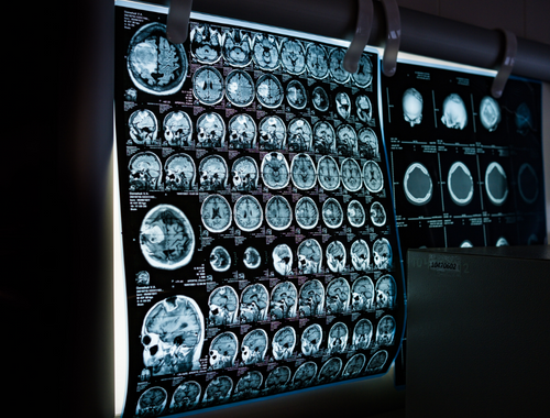
Magnetic resonance imaging (MRI) creates precise, high-resolution cross-section images of the soft tissues and bones of the body using radio waves and a powerful magnet. MRI is typically used to identify bone and joint issues, torn ligaments and cartilage, and herniated discs, and it does not expose the patient to radiation like X-rays, and CT scans do.
During an MRI scan, the patient remains on a table which slides into the tube-shaped MRI scanner. The apparatus then creates a magnetic field around the patient and pulses radio waves into the area of the body that needs to be scanned. The body’s tissues resonate as a result of the radio waves. An advanced computer algorithm translates these vibrations into finely detailed 2D images that are recorded.
The MRI equipment does make a loud noise but is painless, like an X-ray or CT scan. Let your doctor know if you have metal implants, clips, or other body parts because the MRI employs strong magnets.
Similarities between X-ray and MRI
Both Are Used for Diagnostic Purposes
Doctors can identify various injuries, conditions, and diseases with X-ray and MRI scans. X-rays and MRIs are reliable, noninvasive, and safe methods for medical professionals to assess your health.
Doctors Can View the Body’s Internal Structures
X-rays produce images by passing energy beams through bodily tissues onto a specific film. Your interior organs, bones, and tissues are shown in images. On X-rays, bone and metal are visible as white. MRI creates precise images of every internal body part, including the organs, bones, muscles, and blood arteries. As a result, both make it possible for doctors to view the body’s internal structures.
Utilize Various Forms Of Electromagnetic Energy
An X-ray is a diagnostic procedure that creates images of inside organs, bones, and tissues on film using invisible electromagnetic radiation rays. Invisible electromagnetic energy beams are used in X-rays to create images of inside organs, bones, and tissues on film. Additionally, an MRI uses a complex fusion of radio-frequency, low-frequency, and static electromagnetic fields.
FAQ
What are the similarities between MRI and CT scans?
Injections of a contrast dye are frequently used before or during CT and MRI procedures, which enhances the radiologist’s ability to see organs and other body tissues.
What are the similarities between MRI and ultrasound?
MRIs and ultrasounds are excellent tools for allowing doctors to see the inside of the body. Both are painless and outpatient procedures. The imaging tool relies on the area of your body that must be examined. For example, both tools view inside organs and blood arteries.
What are the differences and similarities between MRIs and CT scans?
MRI and CT scans produce images in distinct ways but have similar applications. Although an MRI scan provides a better overall image of the investigated tissue, a CT scan is usually appropriate for bigger areas. In addition, although both procedures have hazards, they are generally safe.
What is the most common MRI?
A functional MRI (FMR), which measures brain activity, a cardiac MRI, which creates detailed images of the heart, and an MRI of the breast are among the most popular types of MRI.
What is the difference between MRIs and X-rays?
MRIs produce a sharper image of soft tissue than CT scans and X-rays. Thus they are frequently used to diagnose problems affecting the brain, muscles, and nerves.
What does an X-ray show that an MRI doesn’t?
X-rays can be used to detect diseased tissue but are more usually used to inspect broken bones. MRIs provide a more accurate evaluation of soft tissues, including brain tumors, spinal cord injuries, and tendon and ligament injuries.


Leave a Reply