What are Archaebacteria and Eubacteria?
Archaebacteria
Archaebacteria, also termed as ‘archaea’ constitute a kingdom of microorganisms having a single cell. The word ‘archaea’ comes from an ancient Greek word ἀρχαῖα meaning ‘ancient things’. These are prokaryotes (organisms without a well-defined nucleus). Archaebacteria and bacteria are thought to be alike in size and structure but scientist discovered that they show radical difference in their molecular organisation due to which they have now earned their own category. These bacteria are not a source of any illness in humans. They contain a distinctive rRNA (Ribosomal ribonucleic acid) i.e. RNA constituent of the ribosome, important for the protein synthesis and this distinctive feature makes them different from their fellow prokaryotes. Archaebacteria play a significant role in biogeochemical cycles like the carbon cycle and nitrogen cycle. The biochemical activity in archaebacteria is diverse and is dependent on various substrates for getting energy and carbon Archaebacteria have been recognised to reproduce asexually occurring by binary fission, fragmentation and even budding.
Archaebacteria are mostly extremophiles as they have the ability to thrive in extreme conditions of acidic environments, extreme saline conditions, high temperatures, pressure or chemical concentration and all these conditions are detrimental to most of the life on planet earth. They are categorised into three main types: crenarchaeota, euryarchaeota and korarchaeota.
Crenarchaeota are the most abundant archaebacteria that account for around 20% of the microorganisms in the form of picoplankton (photosynthesis performing phytoplanktons consisting of cells that range from 0.2 and 2 μm) in the global oceans. These thrive in either very hot (hyperthermophiles), very cold (thermoacidophiles i.e. extremophilic microorganisms that are both thermophilic and acidophilic) and acidic conditions like the hot springs and submarine hydrothermal vents.
Euryarchaeota comprise of halophiles (organisms growing in saline conditions) and methanogens (methane producing bacteria that survives in saline and oxygen lacking environments). For e.g. euyarchaeota resides in the guts of certain animals. Scientists do not know much about korarchaeota except that they thrive in hot springs and are thermophilic.
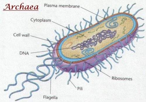
Figure 1. Structure of an archaebacteria cell (Bateman, 1997)
Eubacteria
The Eubacteria, also termed just “bacteria,” belong to kingdom monera. These bacteria lack membranes bound organelles, like mitochondrion’s and chloroplast. All the biochemical activities occur in the cytoplasm. Eubacteria are accountable for several sickness issues in humans and they also constitute significant parts of all of the Earth’s ecological systems. Eubacteria also have a role in nitrogen cycle. They are show parasitic and pathogenic influence on their host life. Apart from asexual methods of reproduction, bacteria reproduce through conjugation as well. Bacteria comprise of a single circular chromosome present in their cytoplasm.
Types of eubacteria include gram positive and gram-negative bacteria. Gram-positive bacteria are the bacterial microorganisms that show positive in the Gram stain test, which enables classification of bacteria into two types on the basis of their cell wall. Gram-positive bacteria potentially take up the crystal violet stain and appear to be purple in colour when seen through the microscope. They appear purple because of the presence of thick peptidoglycan layer in the cell wall absorbs the violet stain after it washes away from the remaining sample, during the decolorization step. Gram-negative bacteria do not possess the ability to absorb violet stain. The reason is that the alcohol used destroys the outer membrane of the gram-negative cells making the cell wall permeable effecting its capability to retain the crystal violet stain. The peptidoglycan layer in gram-negative bacteria is quite thin and appears red or pink as it is present between the inner and outer cell membrane causing the bacteria to take up the counterstain. Gram-positive bacteria are more receptive to antibiotics due to the lack of outer membrane.
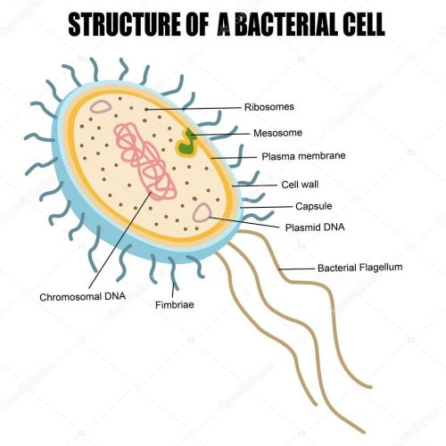
Figure 2. Structure of a bacterial cell (Doetsch & Sjoblad, 1980)
Difference between Archaebacteria and Eubacteria
1. Habitat
Archaebacteria
These consist of organisms that thrive in extreme conditions (temperature as low as 4o C and as high as 120o C) of environment e.g. volcanoes, hot springs, deep ocean vents, salt lakes, marshlands. In human microbiota, they are majorly found in colon, skin membrane and oral cavity. They live in environments that have pH which ranges from 5 to 1. There exist three types of archaebacteria depending upon the habitat preference namely halophiles (microorganisms that thrive in saline conditions), methanogens (bacteria that reduce Carbon dioxide to methane), and thermophiles (microorganisms that survive best at temperatures higher than the normal).
Eubacteria
Eubacteria are present everywhere and are distinct in terms of evolution. They are seen in several habitats like soil, water, in or on the large organisms. These microorganisms are varied and quite adaptable. Eubacteria can be aerobic (which need oxygen) or anaerobic (which do not need oxygen). Some eubacteria even give birth to endospore which helps them to thrive in extreme and harsh environments like freezing, extremely hot, environments with ionizing radiations or particles or chemically wrathful conditions.
2. Structure, size and shape
Archaebacteria
A single archaebacterium ranges between 0.1-15 μm in diameter. Generally, all the archaebacterium except Thermoplasma and Ferroplasma are enveloped by a cell wall which is formed of proteins embedded in the surface layer and is called as S-layer. This layer ensures physical and chemical protection and acts as a barrier for macromolecules to interact with the cell membrane. Archaebacteria do not have peptidoglycan (a polymer comprising of sugars and amino acids which creates a mesh-like protection on the outer side of the plasma membrane for most of the bacteria) inside their cell walls. Archaebacteria are spherical, rod shaped, plate like, spirals, flat and even square in shaped.
Eubacteria
A single individual archaebacterium ranges from 0.1-15 μ m in diameter. The cell wall of archaebacteria does contain pseudo peptidoglycans. The bacterial cell does consist of cell wall and component structures inside and outside the cell wall. Some component structures include capsules mostly composed of polysaccharides. Some capsules are hetero polysaccharides, for example, capsule of Klebsiella pneumoniae and some are polypeptides e.g. anthracis. Capsules play a significant role in disease causing ability of eubacteria.
One more component is the flagella that are thread like, skinny and thin rigid structures, 20 nm wide and around 15 to 20 µ m in length. There is another component structure called Spinae that help bacteria to thrive in harsh environmental conditions like pH, salinity and extreme temperature. Another component is Fimbriae or Pili which are adhesive structures that help bacterial cells to stick to some other bacteria or to a substrate or to some eukaryotic like mould plants and animal cells. Fimbriae is straight and having 7 nm diameter.
Based on cell envelope and Gram stain, bacteria are categorised into two types namely gram-positive bacteria and Gram-negative bacteria. Gram positive bacteria consists of multi-layered cell structure and Gram-negative bacteria contains single type of molecule and is usually quite thick.
Cells of bacteria are varied in their shape and mostly are divided into four conventional shapes namely Cocci (Micrococci, Diplococci, Streptococci, Tetrad, Sarcinae, Staphylococci), rod shaped (short rods and long rods), Vibrios and Spirilla. Some cells ae flat or even square shaped.
3. Membrane Lipids
Archaebacteria
Membrane lipids (double layered surface of all cells) of archaebacteria are unique and distinct from those found in eubacteria. The polar lipids of Archaea are generally utilized to develop liposomes (blocked vesicles) with features that are beneficial in biotechnology. The Archaea consist of membrane lipids composed of hydrocarbon chains with extensions connected to glycerol by ether linkages or branched containing D-glycerol phosphate.
Eubacteria
The bacteria or the eubacteria consist of membranes comprising of unbranched fatty acid chains connected to glycerol phosphates by means of ester linkages, fatty acids with straight chains.
4. Deoxyribonucleic acid (DNA) dependent Ribonucleic acid (RNA) Polymerase
Archaebacteria
Archaebacteria have complex subunit pattern
Eubacteria
They consist of simple RNA polymerases.
5. Antibiotic sensitivity
Archaebacteria
Archaebacteria show no antibiotic sensitivity
Eubacteria
Eubacteria are sensitive to antibiotics.
6. Introns
Archaebacteria
Introns (a section of a Deoxyribonucleic acid or Ribonucleic acid molecule which does not code for proteins and acts as a barrier for the sequential pattern of the genes).
Eubacteria
Introns are absent.
7. First amino acid in all proteins
Archaebacteria
Formylmethionine
Eubacteria
Methionine
8. Growth and reproduction
Archaebacteria
Archaebacteria reproduces by binary fission, fragmentation and budding.
Eubacteria
These also reproduce by binary fission, budding and fragmentation but they are also capable of producing spores which help them to remain dormant in harsh conditions.
9. Glycolysis/Kreb’s cycle
Archaebacteria
Archaebacteria have neither glycolysis nor Kreb’s cycle.
Eubacteria
Eubacteria have both.
10. Examples
Archaebacteria
Halobacterium, Thermoproteus, Thermoproteus tenax, Vulcanisaeta moutnovskia, Sulfolobus islandicus, Ferroplasma
Eubacteria
Bacillus, Anaerobacter, Methanococcus vannielii, Thermococcus sibiricus, Clostridium.
Conclusion
Archaebacteria, eubacteria and cyanobacteria all belong to the kingdom monera. Archaebacteria are also termed as the primitive bacteria whereas the bacteria or the eubacteria are termed as the real or true bacterial organisms. Bacteria or eubacteria are abundantly found in soil, water, live inside and outside of larger species. Bacteria or eubacteria are categorised into two types namely gram positive and gram-negative bacteria. Bacteria which are found in hot springs, volcano vents, volcanoes, water strongly impregnated with salt, deep bed of oceans etc. are the Archaebacteria. Archaebacteria evolved after the birth of first life on this planet. Three categories of archaebacteria can be seen: methanogens, halophiles and thermoacidophiles. The major distinctive characteristic between archaebacteria and eubacteria is their habitation in the ecological system.


Leave a Reply