Doctors routinely request diagnostic imaging tests to monitor what is happening inside your body. The various imaging tests help the doctor make a clear diagnosis and decide on the best treatment option.
Your doctor can identify specific medical conditions with the use of images produced by these imaging tests.
What is a CT Scan?
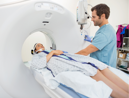
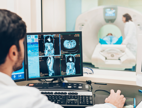
CT, computerized axial tomography, produces images of the body, including bone, using rotating x-rays. The patient lying on the table is rotated around by the x-ray tube. The x-ray detector is located on the patient’s opposite side. This detector picks up the beam that passes through the patient.
The best uses for a CT scan (also called a CAT scan) are evaluating bone damage, identifying lung and chest conditions, and finding cancer.
- Bone structure imaging using CT is effective.
- Some people cannot have an MRI but can have a CT scan if they have specific surgical clips, cardiac monitors, metallic fragments, or pacemakers.
- Compared to MRI, the total testing time is less with CT.
- For claustrophobic people, CT may be more comfortable.
What is an MRI?
A strong magnet and pulsed radio waves are used in MRI (Radio Frequency or RF). Any axis of the body is used to reconstruct the acquired data into a two-dimensional image.
Since bone is devoid of water, it does not produce any picture data. As a result, the photos have a black area. Therefore, the imaging of soft tissue is best suited for MRI scanners.
Doctors can use MRI technology to see soft tissues like muscles, ligaments, tendons, bones, and organs. The spinal cord and nerves can also be seen.
MRI is also used:
- To identify sports injuries such as strained or sprained muscles, torn anterior cruciate ligaments, and ruptured Achilles tendons
- Discover malignant and benign tumors
- Recognize issues with the circulatory, digestive, circulatory, and respiratory systems.
- Check for any abnormalities in the brain, such as aneurysms and tumors
- Identify bone and cartilage diseases
- See how much your joints are inflamed.
Similarities
Noninvasive
The noninvasive diagnostic imaging process known as computed tomography (CT scan or CAT scan) creates horizontal or axial images of the body (commonly referred to as slices) using a mix of X-rays and computer technology. The CT scan is a noninvasive, pain-free, and relatively safe process that doesn’t require recovery time.
The noninvasive medical imaging procedure known as magnetic resonance imaging, or MRI, creates precise images of every internal bodily structure, including the organs, bones, muscles, and blood arteries. MRI scanners create images of the body by using a powerful magnet and radio waves.
Detailed Images
Soft tissue, bone, and blood vessel details can all be seen in great detail in CT scans. MRI scans are better and more accurate at visualizing the ligaments, soft tissue, or organs.
Soft tissue injury, ligament damage, and herniated discs may be simpler to detect as issues after an MRI scan. Medical professionals can use a CT scan to obtain images of organs, fractured heads, or physical components.
The Machine
In a CT scan and an MRI scan, the patient is on a bed that gently rotates across the gantry as a narrow beam of x-rays is shot into the body by the x-ray tube in case of a CT scan. For MRI, the magnetic field momentarily realigns your body’s water molecules. Then, radio waves induce these aligned atoms to emit weak signals to make cross-sectional MRI pictures.
You must lie still and motionless throughout the scan. If you move, the MRI or CT scan images might need to be clarified. If you are claustrophobic (afraid of enclosed places), have trouble staying still, or suffer chronic pain, your primary care doctor might prescribe you a light sedative.
FAQs
Are CT and MRI contrast the same?
MRI and CT use the same contrast materials, except iodine, which is only present in CT.
What are the differences between MRI and FMRI?
MRI and fMRI scan both employ the same fundamental atomic physics concepts. However, MRI scans show anatomical image structure, and fMRI scans show metabolic function. As a result, MRI scan results resemble three-dimensional photographs of anatomical structures.
What can an MRI show that a CT scan cannot?
A CT scan cannot detect some disorders, which is where MRI excels. Some tumors, including liver cancers, uterine cancers, and prostate cancers, are virtually undetectable or extremely difficult to find on a CT scan. An MRI is also better at showing bone and brain metastases.
Which is safer, MRI or CT scan?
A tiny dosage of ionizing radiation is used in CT scans to create the images. However, an MRI scan doesn’t operate in this manner. Instead of ionizing radiation, it creates images using strong magnets and radio waves. So, in contrast to a CT scan or x-ray, you are not exposed to radiation when you undergo an MRI scan although it does take longer.
Why would a doctor order a CT scan instead of an MRI?
The doctor might order a CT scan instead of an MRI if a patient cannot have an MRI. Due to the strong magnet inside the machine, those with metal implants, pacemakers, or other implanted devices shouldn’t undergo an MRI.
Is MRI more expensive than CT?
MRIs are substantially more expensive than CT scans and other imaging procedures due to the higher equipment expenses. Additionally, reading these intricate images by radiologists requires additional time.
Why do they inject dye for an MRI?
Dye is injected because MRI scans sometimes require it as a contrast agent. Certain tissues and blood arteries become more distinct and detailed as a result.
Can you do CT and MRI at the same time?
CT and MRI data are spatially and temporally registered in a perfect CT-MRI scanner. Therefore, CT and MRI scans that were collected separately could be combined to mimic a simultaneous acquisition.


Leave a Reply